
Magic Salamanders & the Zero-Point Field (1 of 5)
The Principles of Regeneration

The ability to regenerate an injured or amputated limb is considered mythological by human standards. The famous book, “Autobiography of a Yogi” cites an incident in which a roadside beggar purportedly regenerated his severed arm. In the fictional comic book series “X-men”, the character known as Wolverine has special regenerative capabilities. If a human could reliably demonstrate the dramatic ability of limb regeneration in this modern age, they would be considered superhuman beyond superhuman. The amusing thing is that there is a vertebrate with 2 eyes, 4 limbs, that walks on land, swims in water, and has virtually the same internal organs of a human that can regenerate their severed limbs. We do not consider this animal to be supernatural in any sense of the word. We just merely refer to this creature as “salamander”. This animal has the ability to not only regenerate their limbs and tail… they have also been documented to regenerate their brain, heart, liver, lungs, spinal cord, teeth, eye lenses, and ovaries (Bölük et al., 2022). They truly are regenerative machines seemingly having the ability to regenerate nearly an entire specimen. The average human by contrast is largely known to be relegated to solely being capable of regenerating their skin and liver cells.
One of the most important books published within the past 100 years is titled, “The Body Electric: Electromagnetism and the Foundation of Life”, authored by Dr. Robert O. Becker. The book is an intriguing tale of a medical doctor who did basic regenerative research while also applying his knowledge to individuals at the VA Medical Center in Syracuse, New York. It’s a rare glimpse of an inside look to the thought process of a brilliant researcher who was able to investigate the inner workings of regeneration in salamanders while applying some basic principles to help patients. Instead of primarily focusing on the genetic and biochemical aspects of this ability, Dr. Becker focuses on the bioelectric changes that take place during the process of regeneration. He also provides a glimpse as to the changes between organisms that have this ability (salamanders) and those that do not (frogs, rats, humans).

The intrigue of the salamander is based on the fact that unlike other regenerative animals such as worms, starfish, and crustaceans, the salamander is anatomically similar to humans.
The question is… how do they do it?
It’s still an incomplete puzzle… hence the name, “The Magic Salamander”.
Nevertheless let us attempt to deconstruct some aspects of this magical situation.
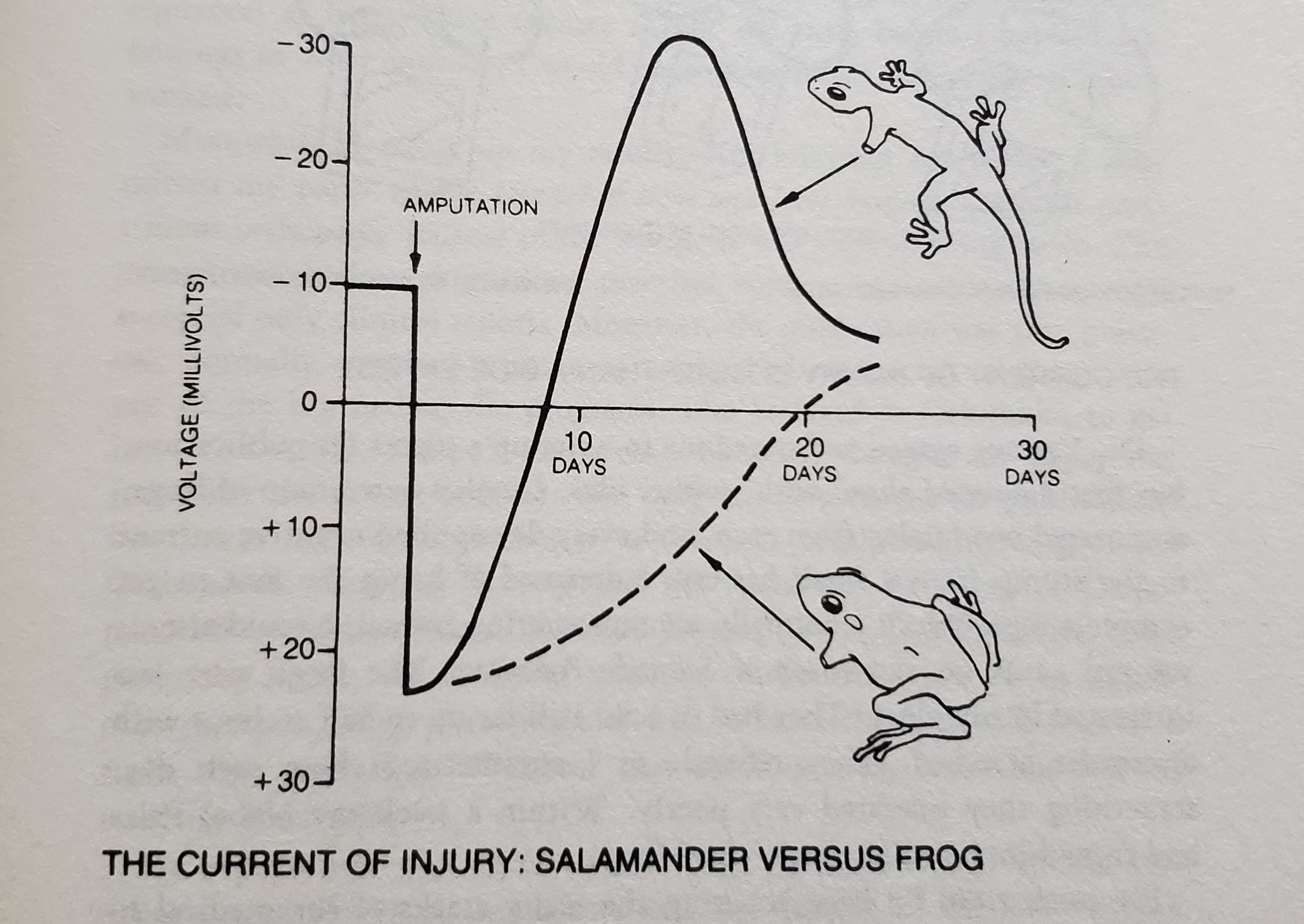
(An image on page 73 illustrating the differences between the direct current (DC) voltage changes between the Magic Salamander that can regenerate their limbs and the lowly frog incapable of doing so.)
As shown above, there is a distinct difference in the autonomic response regarding the bioelectric signaling of wound healing and limb regeneration between the two species. Let us present an excerpt from “The Body Electric”. (For reference the term blastema refers to a mass of cells capable of growth and regeneration.):
“I made measurements daily, expecting to see the salamander voltages climb above those of the frogs as the blastemas formed. It didn’t work that way. The force of the current flowing from the salamanders’ amputation sites rapidly dropped, while that from the frogs’ stumps stayed at the original level. By the third day the salamanders showed no current at all, and their blastemas hadn’t even begun to appear. The experiment seemed a failure. I almost quit right there, but something made me keep on measuring. I guess I thought it would be good practice. Then, between the sixth and tenth days an exciting trend emerged. The salamander potentials changed their sign again, exceeding their normal voltage and reaching a peak of more than 30 millivolts negative just when the blastemas were emerging. The frogs were still plugging away with slowly declining positive voltages. As the salamander limbs regenerated and the frog stumps healed over with skin and scar tissue, both groups of limbs gradually returned (from opposite directions) to the original baseline of 10 millivolts negative.”
This was an extremely important finding from Dr. Becker regarding not only the differences between species but also a distinct shift from his initial hypothesis of a linear reaction taking place. When a limb is fully intact, in a normal setting the measurement sits at a baseline of -10 millivolts (mv) negative. When a wound or amputation occurs, the direction of the current quickly reverses towards a positive polarity reaching over 20 mv (positive). Becker originally expected the regenerative aspects of the salamander to coincide with a continuation of the positive polarity exceeding that of a frog (20 mv) believing that this projected increase coincided with the regenerative process. This was not the case and instead new insights developed from this observation.
Harold Saxton Burr was a Yale professor from 1929 to 1958 with a focus on neuroanatomy and bioelectrodynamics. He would eventually author a book summarizing his findings titled, “Blueprint for Immortality: The Electric Patterns of Life”. Burr would spend much of his career measuring electric fields and electric potentials on the surfaces of a wide array of organisms including worms, salamanders, humans, other mammals, and even slime molds. His lab would measure the changes in the electric potentials and their correlations with growth, regeneration, tumor formation, drug effects, hypnosis, and sleep. One of the most significant data points that Burr generated was observing dipolar distribution of potentials in most animals with their heads being positive and their tails being negative. Dipolar distribution can be described as equal or opposite electric charges separated at a distance. Becker stated that while Burr’s work was important, the equipment utilized for his experiments were too “noisy” to reliably measure the minute currents found in living things.
Becker would attempt to replicate Burr’s findings employing more modern equipment on salamanders. What he found was a more complex field that followed the arrangement of the nervous system. Becker observed that there were large positive potentials over the brain and slightly smaller ones over the spine in between the limbs. Instead of simply finding a negative charge at the tail, he found negative charges at the tip of the nose, the feet, hands, and the tip of the tail. Additional organisms such as flatworms, earthworms, fish, amphibians, reptiles, mammals, and humans were all tested as well.
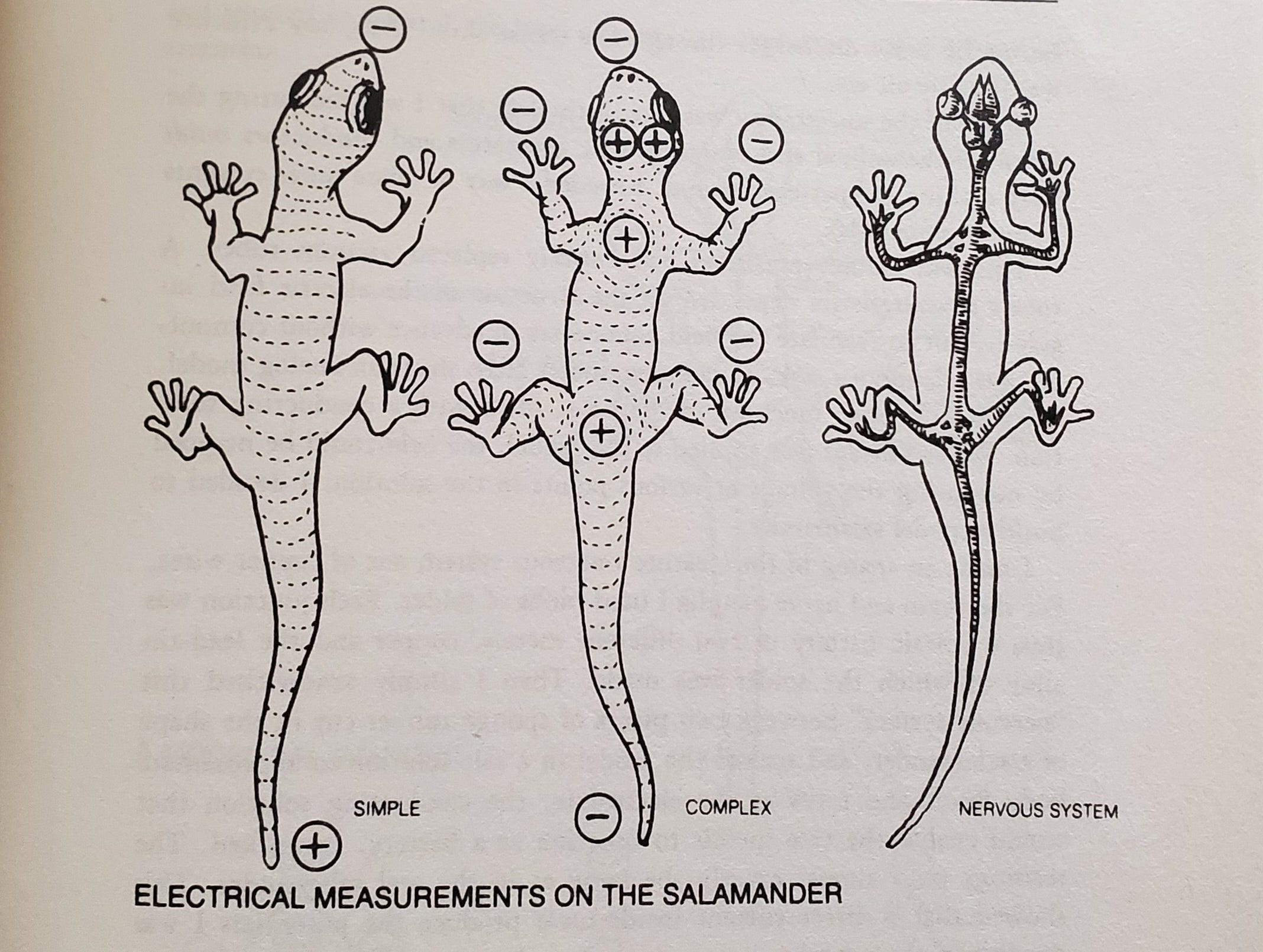
(An image on page 95 comparing the simple body electric field from Burr compared to the complex body electric field from Becker.)

(Images from pages 97 & 98 showing the direct current (DC) pattern in various species.)
While this is all mildly interesting information in a vacuum, the application of this knowledge is what is increasingly intriguing. In 1960’s, a researcher named Stephen D. Smith at the University of Kentucky was studying the effects of electrical stimulation on limb regeneration in frogs. Smith would implant a tiny battery with silver and platinum wires at the site of amputation of a frog’s arm. The effects were tested by bending the wires into the bone marrow cavity with some frogs having the silver wire emitting a positive current into the marrow and others having the platinum wire emitting a negative current into the marrow. What Smith found was that the limbs with the positive silver electrodes showed no growth, and in some cases showed tissue disintegration. By contrast, the limbs with the negative platinum electrodes showed the start of the regenerative process. These results concur with the findings of Becker in the first chart up above (p. 73) in which the differentiation between the salamander regenerative abilities and the frog’s inability is based on the negative current strength following amputation. By 1974, Smith had refined his electric application device to the point of achieving full regrowth of a frog limb (Smith, 1974).
In 1971, based on the ongoing work from Smith, Dr. Becker would attempt to apply this same electric stimulation to rats who had their limbs amputated. Leading up to the study, he was skeptical of whether this would be possible being that the proportion of nerves to other tissues in rat legs was 80 percent less than in salamander legs. Nevertheless, Becker proceeded with the study utilizing a medium-current device supplying 1 nanoamp with silver and platinum wires at the site of the rat limb amputation. Following 1 week of the electric implant, the experimental rats had a well-formed blastema with ten different kinds of cells differentiating out from it. Being that the amputation had occurred above the elbow, some of the animals showed two cartilaginous deposits appearing to be the precursors of the two lower arm bones. In one of the animals that had experienced the electric stimulation for a longer period of time, cartilage in a five-fingered shape was observed indicating that a hand had begun to grow (Becker et al., 1972).
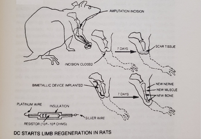
(Illustration from page 153 showing the process of externally stimulated electric signal for limb regeneration in a rat.)
While this initial experiment by Becker did not showcase full regeneration of an adult rat limb, it showed enough complex differentiation for follow up researchers to verify and replicate the effects (Person et al., 1979; Leppik et al., 2015; Oliveira et al., 2019). One of the leading research groups doing extensive work in the field of bioelectricity today is the Levin Lab at Tufts University. In 2011, this lab published a study observing for the first time that a complex bioelectric signal preceded the cellular formation of a frog face. One of the lead researchers of that study, Dany S. Adams would serendipitously come across this development by leaving a microscope camera on overnight while imaging the development of frog embryos. Adams didn’t believe that the video quality would be clear enough to capture anything of importance based on the movements of the embryo. However, the next morning she checked the video, saw that the images came out clear, and as they say… the rest is history. One potential impact point of this finding is that it adds support to the concept of the morphic resonance and morphic field theory with possible interactions with electricity as a top-down driver of this model. The concept of morphic resonance/field theory is that it is based on the notion that there is a field that influences biological development carrying with it the information needed to organize and stabilize the physical form.
Perhaps electricity is key in this equation?
How does any of this tie into the human potential?
An interesting phenomena that has been documented for centuries is the ability for humans to shut off the pain response when in a state of trance. The forbidden word for trance is known as “hypnosis”. While there is much bias against the term based on the popularized idea that stage hypnotists make people do strange things… there is undoubtedly a significant body of legitimate research in the field of clinical hypnotherapy. There are over 16,000 search results regarding hypnosis & hypnotherapy in the peer-reviewed literature on pubmed.
The extraction of the third molar (wisdom tooth) is a procedure that normally requires general chemical anesthesia. This is the process of essentially putting a person to sleep so that they do not feel the pain of the tooth removal. Most people cannot fathom undergoing this process without chemical anesthesia of some kind. However, there are subjects that are allergic to chemical anesthetics and therefore do not have the option to utilize these substances for surgical procedures. For these patients, their only alternative is to either simply bear the pain of surgery as is or rely on hypnosis to provide an analgesic effect. There are numerous case studies of subjects undergoing this exact procedure of teeth removal while solely utilizing hypnosis induced analgesia (Abdeshahi et al., 2013; Cozzolino et al., 2020; Facco et al., 2021; Shaw et al., 1996; Glaesmer et al., 2015; Ghoneim et al., 2000; Schmierer et al., 2023; Queirolo et al., 2024; Sabherwal et al., 2021). For people skeptical of the peer-reviewed literature of such a phenomena, a clinical hypnotherapist and friend of ours Sharon Waxkirsh has documented this procedure in video format. There is even footage of Sharon utilizing self-hypnosis on herself to undergo wisdom tooth extraction without chemical anesthesia.
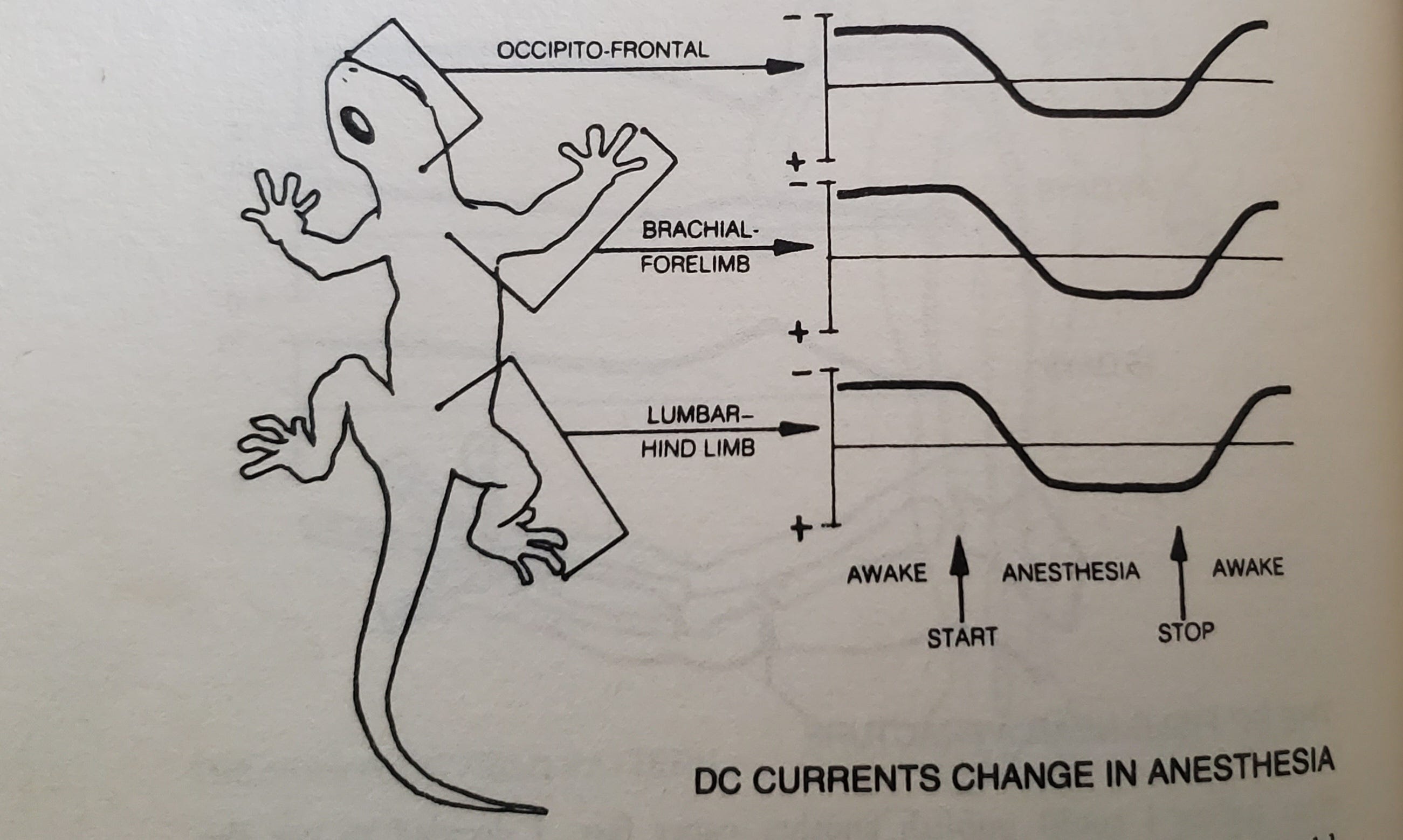
Interestingly, one of the skeptics of hypnosis was Dr. Becker. Here is an excerpt from The Body Electric regarding his research into hypnosis, pain control, and bioelectric changes:
“Charlie, Howard, and I decided to find out how the brain’s DC potential behaved in humans. The electrodes we’d been using on salamanders couldn’t be scaled up for people, but within a week Charlie invented some that would give us equally precise readings from the human head. We immediately found that the back-to-front current varied with changes in consciousness just as in salamanders. It was strongest during heightened physical or mental activity, it declined during rest, and it reversed direction in both normal sleep and anesthesia. This knowledge led directly to the experiments, described in Chapter 13, that taught us much about how hypnosis and pain perception work.” p. 116
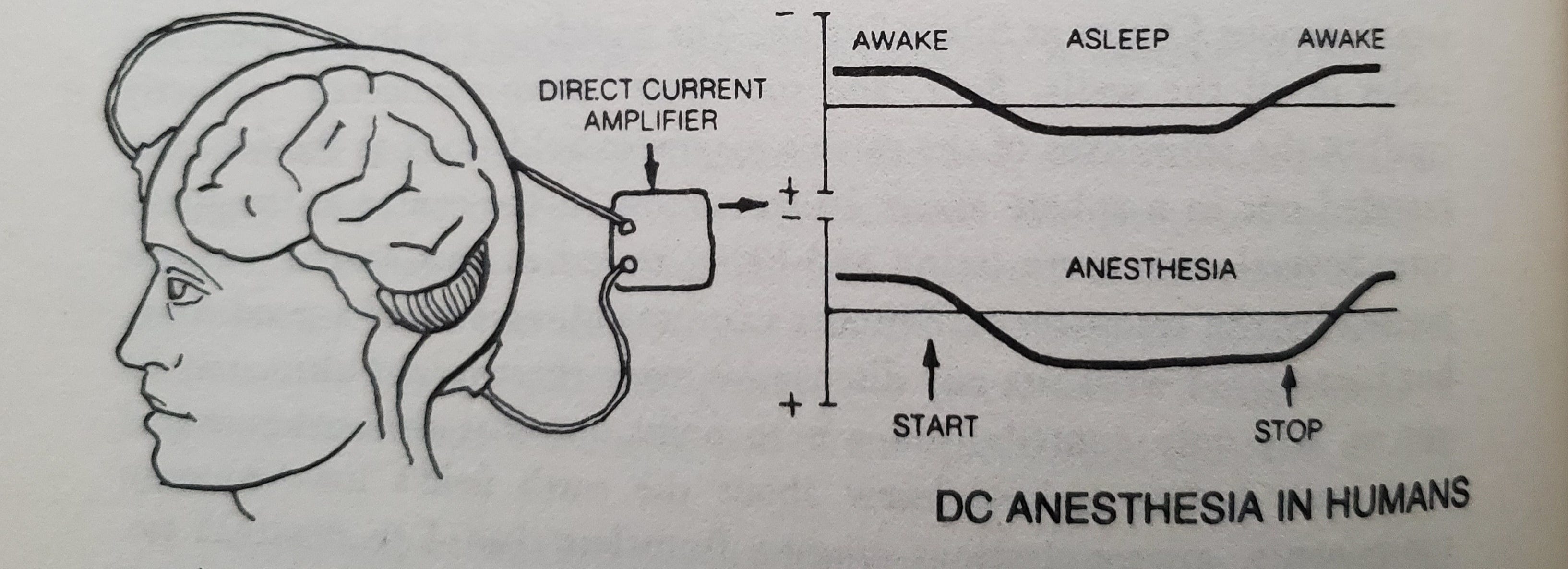
(Image from page 116 describing the changes in DC current direction based on changes in consciousness.)
“One of the most exciting results of my collaboration with Dr. Friedman was proof that one’s state of waking consciousness could change the perception of pain. Friedman, who already used hypnosis to control chronic pain in his patients, gave several of his best subjects hypnotic suggestions of arm numbness deep enough that they couldn’t feel the prick of a needle. In each case, I found that the frontal negative potential of the head became less negative, often reaching zero, as the client attained deep trance. The reading changed in the same direction as in anesthesia, only not as far. Then, when the suggestion for pain control was given, the arm potential reversed just as it had in response to procaine. Conversely, when a control subject was asked in normal waking consciousness to concentrate forcefully on one arm, its sensitivity to pain increased, and the hand potential became more negative. We found we could use this difference to determine whether a person was really hypnotized or just cooperating.” p. 238
“Some doubters (including myself, I’m afraid) had believed hypnoanalgesia was merely a state in which the patient still felt the pain but didn’t respond to it, but these experiments proved it was a real blockage of pain perception. It seems that the brain can shut off pain by altering the direct-current potentials in the rest of the body “at will”.” p. 239
(Image from page 100 showing the systemic changes coinciding with changes in consciousness. Negative potentials in the brain’s frontal area and at the periphery were associated with wakefulness and sensory stimuli. The systemic shifts toward positive potentials occurred during rest and even more so during sleep.)
Now we are getting somewhere.
It appears that changes in consciousness coincide with changes in the direction and strength of the electric potentials in the brain and body. The changes coincide with the perception of pain in humans under hypnosis. They also seem to change coinciding with the regenerative process in salamanders. Becker cites the following on page 238: “In addition, the DC potentials react slowly enough to account for pain. A wound usually doesn’t start to hurt in earnest until minutes, or even hours, after the injury. This delay has been especially hard to explain in terms of nerve impulses, which travel at 30 feet per second. However, when Friedman and I injured the limbs of salamanders while monitoring the potentials on their limbs and heads, we found that the change in the limb reading showed up in the head after a time approximating that of delayed pain.”
This gives us a signal that there lies the potential that mammalian regeneration is a possibility by utilizing altered states of consciousness to induce the desired results. We understand that the pathway to accomplish and it’s relationship to the Zero-point Field (ZPF) is not quite clear to you as a reader right now but we’ve bombarded you with enough strange information thus far so let us continue clarifying this concept in Part 2 to be released next week.
Stay tuned!